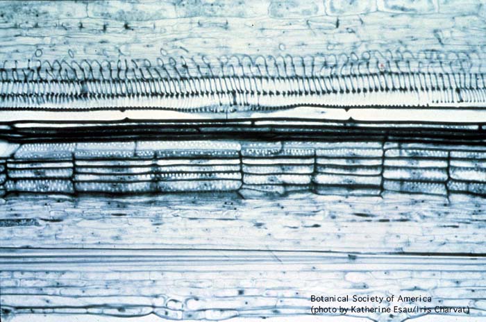
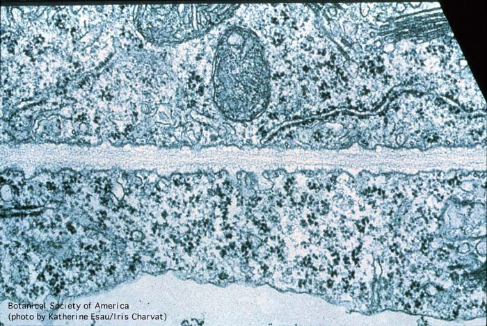
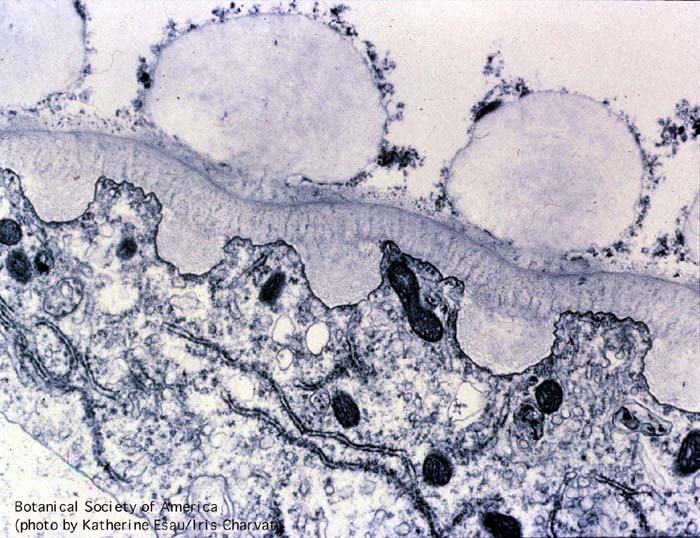
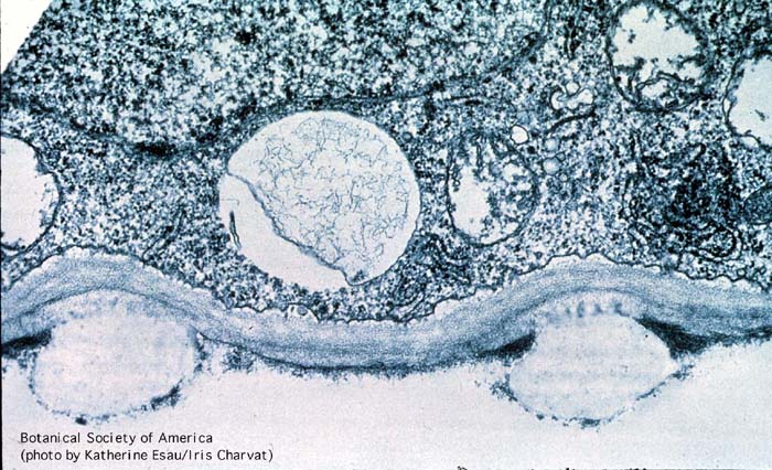
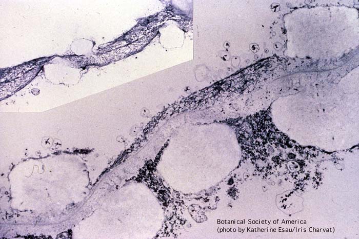
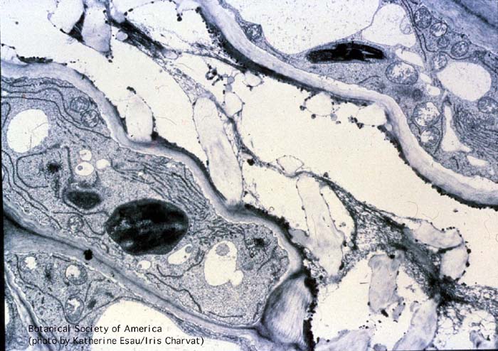
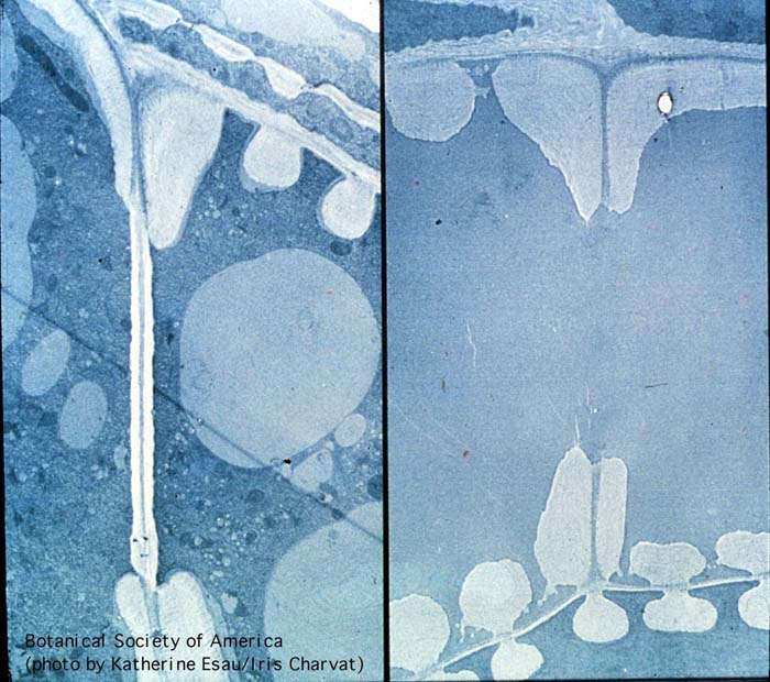
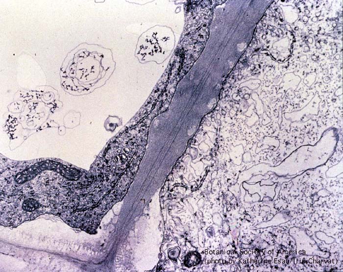
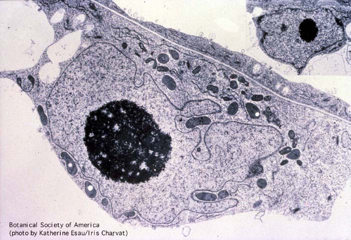
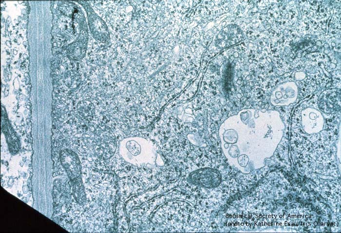
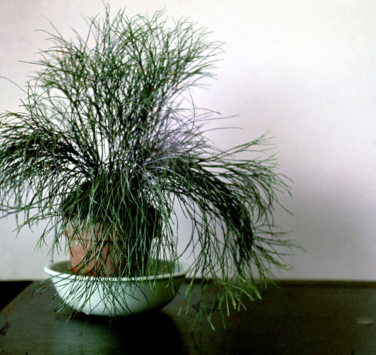
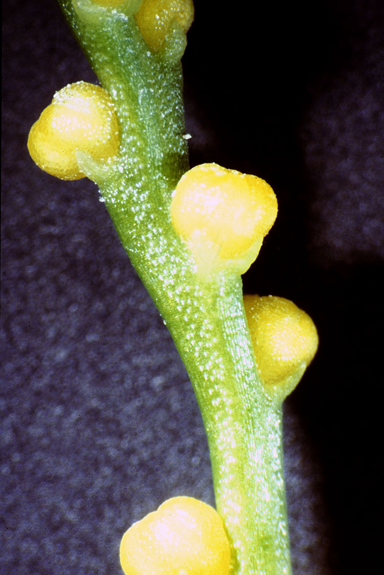
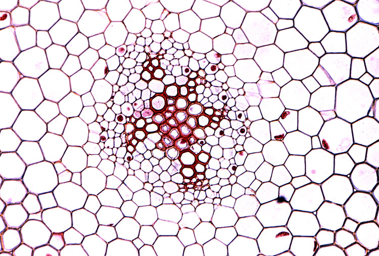
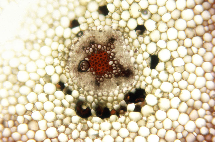
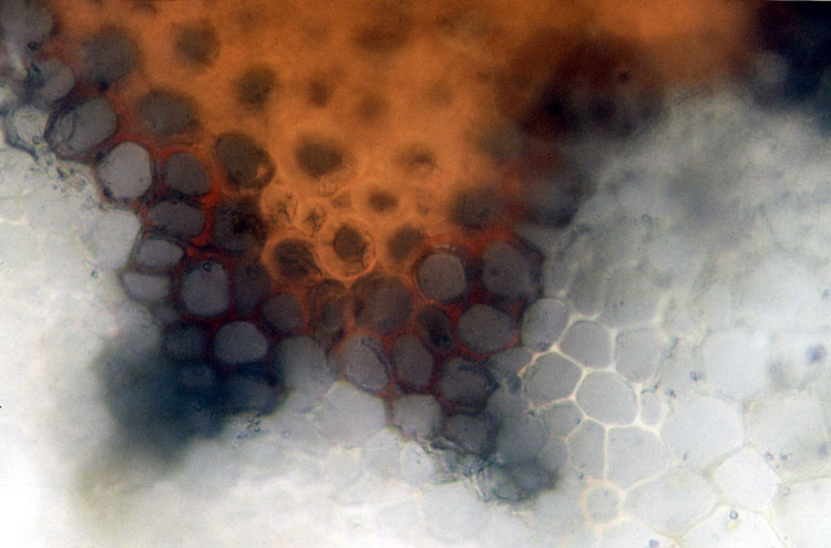
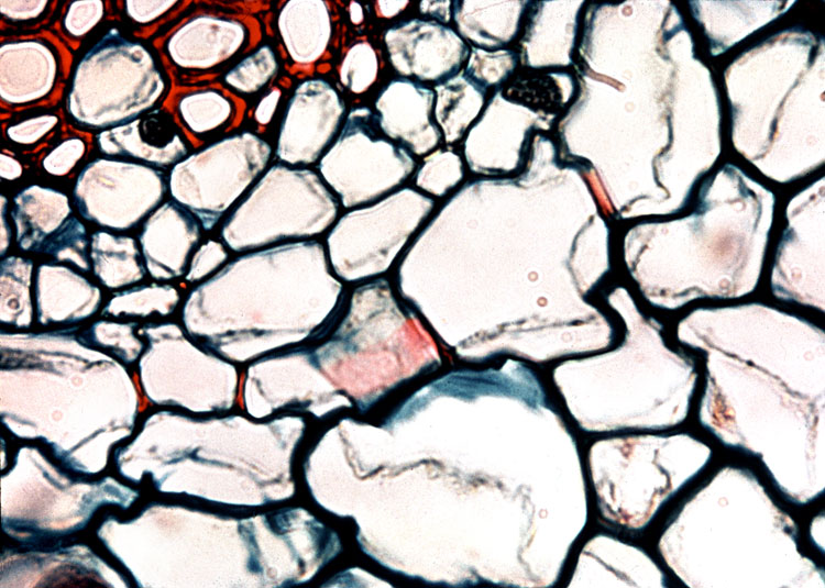
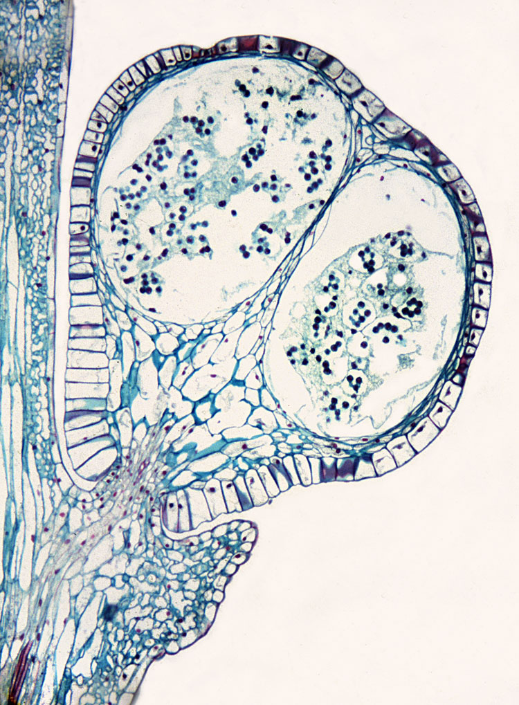
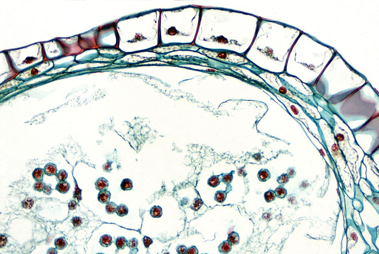
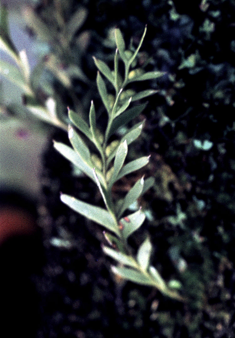
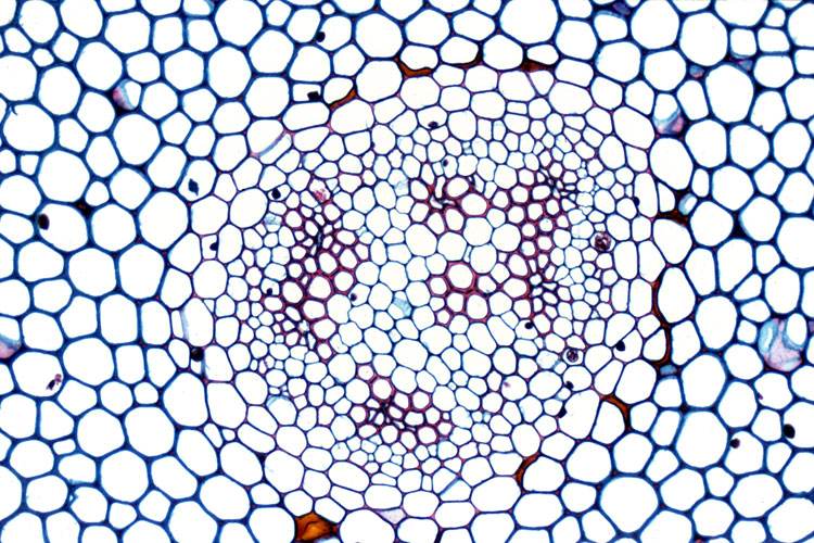
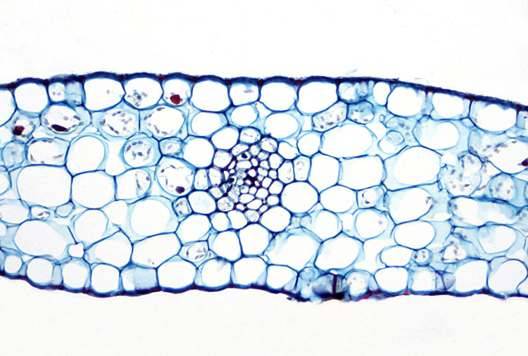
Plant Image Collection

Xylem DevelopmentActions

Media

04-001h Quick view
04-001h

Section through xylem region of a vascular bundle drawn from electron micrographs. Parenchyma cells present; all other cells are tracheary elements in various developmental stages. Scale line, 10 um.

04-002h Quick view
04-002h

Ontogenetic sequence of primary xylem elements (from bottom to top) with annular secondary wall thickenings (rings) gradating to helical thickenings in a moderately stretched cell.

04-003h Quick view
04-003h

Side wall between two differentiating vessel members. Fibrous material is present in vesicles connected with the near wall. Middle lamella and connections between microtubules and plasmalemma.

04-004h Quick view
04-004h

Side wall between differentiating vessel members. Secondary wall deposition not yet evident. In the older cell (below), microtubules concentrate near young helical thickenings and are absent along the primary wall.

04-005h Quick view
04-005h

Side wall between vessel members with differentiating helical thickenings. More mature cell (right) indicated by larger amount of secondary wall deposition. Vesicles between plasmalemma and secondary wall are evident.

04-006h Quick view
04-006h

Wall between older (right) and younger (left) vessel members. Microtubules accumulate near the secondary wall in younger cell. Membrane-bound vesicles appear as blebs on plasmalemma. Primary wall of older cell are partly hydrolyzed.

04-007h Quick view
04-007h

Vessel member above, parenchyma cell below. Primary wall is partly hydrolyzed in the vessel member. Protoplasmic residue covers the secondary and hydrolyzed primary wall.

04-008h Quick view
04-008h

Degradation of primary wall of vessenl member is almost complete. Primary wall of other cell is partly hydrolyzed between the coils of the helix. Older helix subjected to greater amount of stretching.

04-009h Quick view
04-009h

Later stage of end wall degradation.

04-010h Quick view
04-010h

Rim of secondary wall material (top) has been deposited around the margin of the end wall delimiting the future perforation. Part of the end wall not covered by the rim (bottom) has been removed.

04-011h Quick view
04-011h

Vesicles occur near the surface of the exposed wall part and some also within the immature rim. Microtubules near the rim at arrowheads.

04-012h Quick view
04-012h

Nucleus in a xylem parenchyma cell. At cw, primary cell wall between two parenchyma cells.

04-013h Quick view
04-013h

Protoplast of differentiating vessel member with part of end wall. Details in the protoplast include dictyosome with vesicles, mitochondria, microbodies, rough and smooth ER, and autophagic vacuoles.

25-001 Quick view
25-001

Habit view of Psilotum sporophyte

25-002 Quick view
25-002

Close-up view of fertile branch

25-003 Quick view
25-003

Psilotum aerial stem, transverse section (x.s.)

25-004 Quick view
25-004

Psilotum aerial stem free-hand section, phloroglucinol + HCL staining

25-005 Quick view
25-005

Close-up of stele showing fibers in

25-006 Quick view
25-006

Psilotum stem x.s.

25-007 Quick view
25-007

Psilotum synangium l.s.; sporocyte stage; note wispy tapetum, multiple layers of eusporangium wall, foliar appendage

25-008 Quick view
25-008

Psilotum synangium l.s. higher magnification; sporocytes prior to completion of meiosis

25-009 Quick view
25-009

Psilotum gametophyte with anheridium and archegonium; endophytic fungus present in ground tissue of gametophyte

25-010 Quick view
25-010

Tmesipteris

25-011 Quick view
25-011

Tmesipteris aerial stem x.s.; siphonostele, mesarch primary xylem; locate proto- and metaxylem

25-012 Quick view
25-012

Tmesipteris synangium x.s. pre-meiosis stage; note multiple wall layers

Other Collections

This collection is within

Previous, in Botany & Plant Science

Next, in Botany & Plant Science

All, in Botany & Plant Science

Creation and update

Creation
Created by admin on Oct 23, 2013
Last update
Updated by admin on Oct 23, 2013

Back to overview