Plant Image Collection

Physiology, Biochemistry, and CellActions

Media

abot79-7 Quick view
abot79-7

Autoradiograph of a small area of longisection of henbane anther following in situ hybridization with 35s-labeled rice histone antisense probe. Autoradiographic silver grains are localized in epidermis and endodermis (at bottom).

abot80-11 Quick view
abot80-11

Salt-secreting stem cuttings of Avicennia germinans (left) and Frankenia grandifolia (right) placed in a flask containing 30 mM NaCI under continuous fluorescent light. A. germinans salt deposits are scattered white spots, mainly on the upper (adaxial) leaf surface. Long, hairlike, white salt deposits are obvious on both surfaces of leaves and on the internodes of the stem of F. grandifolia.

abot81-2 Quick view
abot81-2

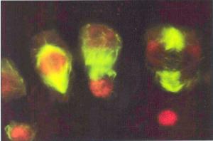
Different stages of cell division in marginal cells of the discoid thallus of the charophyte, Coleochaete orbicularis, double stained to show microtubules (green) and nuclei (red).

abot81-4 Quick view
abot81-4

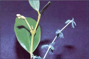
Photograph of a tissue print from a mature leaf of Peperomia camptotricha. A thich hand-cut fresh leaf section was pressed onto a nitrocellulose membrance. The resulting untouched print was photographed with side illumination to give a 3-D appearance. Such prints can be used for histochemical studies.

Other Collections

This collection is within

Previous, in Botany & Plant Science

Next, in Botany & Plant Science

All, in Botany & Plant Science

Creation and update

Creation
Created by admin on Oct 23, 2013
Last update
Updated by admin on Oct 23, 2013

Back to overview